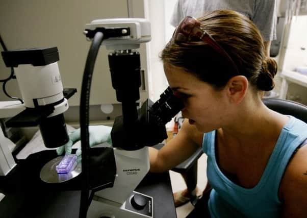
New test could detect cancer within hours


Current tests to look for cancer and other diseases in tissue samples can take weeks to be fully analysed by specialists.
But UK researchers now believe a detailed diagnosis – including information on the subtype of cancer a patient has – could be available in hours using mass spectrometry imaging (MSI), a form of chemical analysis.
Advertisement
Hide AdAdvertisement
Hide AdThe technique was outlined in the journal Proceedings of the National Academy of Sciences.
MSI produces images of the distribution of chemical components in a sample and is already used in fingerprint analysis and archaeological investigations.
Scientists from Imperial College London found that it could also be used to analyse tissue samples to build up a database that would allow rapid diagnosis of cancers and other diseases.
They used a form of MSI which involves a beam of electrically charged solvent droplets moving across the surface of a sample. A machine then processes the information gathered from this to produce a pixelated image. Each pixel contains data on the thousands of chemicals present in that part of the sample.
The researchers said that by analysing many samples in this way, and then comparing them to the results from traditional tissue analysis, a computer could learn to identify different types of tissue.
They said that this could mean that a single test taking a few hours could provide much more detailed information than standard testing.
For example, it could show not only that a tissue was cancerous but also the type and subtype of cancer.
This information is particularly valuable in choosing the best treatment for the patient as more and more drugs are tailored to specific types of cancer.
Advertisement
Hide AdAdvertisement
Hide AdThe scientists said the technology could also be applied in research to give new insights into the biology of cancer.
Dr Kirill Veselkov, from Imperial’s department of surgery and cancer, said: “This work overcomes some of the obstacles to translating MSI’s potential into the clinic.
“It’s the first step towards creating the next generation of fully automated [tissue] analysis.”
Co-researcher Dr Zoltan Takats said the technology could “change the fundamental paradigm of histology [the study of the microscopic anatomy of cells]”.
“Instead of defining tissue types by their structure, we can define them by their chemical composition,” he said.
“This method is independent of the user – it’s based on numerical data, rather than a specialist’s eyes – and it can tell you much more in one test than histology can show in many tests.”
Professor Jeremy Nicholson, head of Imperial’s Department of Surgery and Cancer, said there had been relatively few major changes in the way scientists studied tissue samples since the late 19th century, when staining techniques were used to show tissue structures.
He said: “Multivariate chemical imaging that can sense abnormal tissue chemistry in one clean sweep offers a transformative opportunity in terms of diagnostic range, speed and cost, which is likely to impact on future pathology services.”
Advertisement
Hide AdAdvertisement
Hide AdDr Kat Arney, Cancer Research UK’s science information manager, said: “This research shows how this imaging technique might be applied to tumours, but there’s a very long way to go to turn it into a device or test that could analyse samples from cancer patients on a routine basis.”