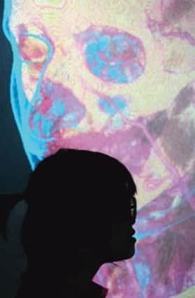
Technology turns patient data into 3D clinical images


Now, technology pioneered at Aberdeen Univeristy to turn real-life patient data into 3D clinical images for the lecture hall has proved so successful, that it is being used as a core teaching method for medical undergraduates.
Professor Alan Denison, deputy head of the division of medical and dental education at the University of Aberdeen, said the technology had helped take teaching of what lies “under the skin” to a new level.
Advertisement
Hide AdAdvertisement
Hide AdAberdeen University is one of the first to use the technology on such a scale.
He said: “This method is really crucial to providiing effective patient care
“Patients of course don’t present in two dimensions, they present in three dimensions.
“Anatomy is a difficult subject to learn. It may be better to hold a bone in your hand but these scans can enrich the learing and understand the complexities of what is going on under the skin
“We stil use cadavars in elements of teaching but patients don’t present as cadavars, they are real people.”
He added: “Blood supply, for example, can be difficult to understand, particularly in the brain, and that is one of a series of packages were have developed.”
The images are created by taking real patient data from CT and ultrasound scans carried out at NHS Grampian hospitals and anonymising the results.
They are then converted into the 3D images, which can be beamed into a special 3D theatre used by both medics and Aberdeen University students.
Advertisement
Hide AdAdvertisement
Hide AdProfessor Denison added: “The students can use iPads to manipulate the anatomy, whether by putting skin on or taking it off the body part of by looking inside the vessels. They can flydown a windpipe or look inside a spinal colum.”
“It is an immersive learning experience and it fun,and learning should be fun.
“We have done a lot of work evaluating the 3D clincial teaching and we found that it has really enriched the student experience.
“We are really using the technoloy of today to train the doctors of tomorrow.”
Professor Denison said there were no plans to phase out the use of cadavars, unlike some medical schools, which he said were still a useful training tool.
He added: “The experience of working with human tissue is an important development in the journey of becoming a doctor and helps to devlop compassion, care and respect of patients.”