Revolutionary microscope brings new focus to fight against disease


The three-dimensional images can be produced within seconds, rather than hours, which could speed up developments of new medicines, said scientists at the University of Strathclyde.
The Mesolens – the only such device in the world – can offer a detailed glimpse of organisms too big for traditional microscopes, as well as allowing imaging of cancerous tissues and the cortex of the brain.
Advertisement
Hide AdAdvertisement
Hide AdThe lens can produce resolution of more than 150 megapixels, equivalent to ten modern digital cameras combined.
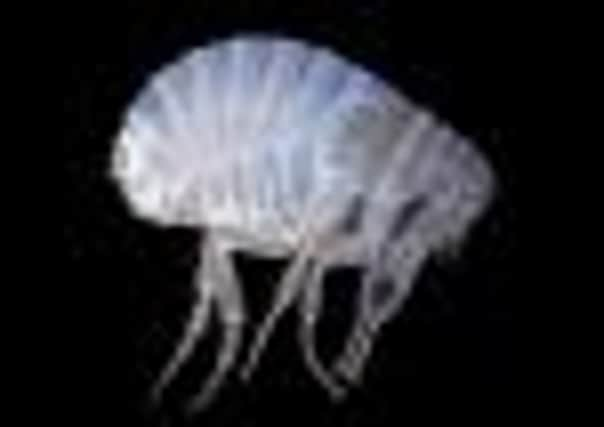
On the 350th anniversary of the Royal Society in 2010, the Mesolens image of the human flea was produced to contrast with the 1665 engraving of a flea by Robert Hooke.
The new lens revealed the buried antennae of the flea, which were invisible to Hooke, whose engraving was otherwise correct.
Dr Amos said: “This level of detail can open up vast possibilities for discoveries.”
Dr Gail McConnell, a reader at the Strathclyde Institute of Pharmacy and Biomedical Sciences, is a partner in the research. She is working with Dr Brad Amos, a visiting scientist at the institute who first conceived the Mesolens project, and Dr John Dempster, who has contributed the software and electronics expertise necessary.
The lens is much larger than a normal microscope lens: about 70cm long and 8cm in diameter. A laser is fired through the lens and scanned over the object, making it emit fluorescent light.
Images from different levels of focus are reconstructed into a three-dimensional picture showing a complete object, but also its interior. The resulting picture resembles a nuclear magnetic resonance scan, but the detail is a thousand times finer.
The new picture contains both the macro information (like the 350-year-old engraving, pictured above, right) and microscopic detail of the interior of individual cells.
Advertisement
Hide AdAdvertisement
Hide AdDr McConnell said it was this combination of macro and micro that is “totally novel: a world first”.
She added: “The Strathclyde project is expected to change the practice of microscopy world-wide, because it closes a serious gap between low and high magnification recording.
“We’re using light to look at biological tissue and understand how it functions. That will assist greatly in helping develop new drugs for an unfortunately infinite number of diseases.
“Currently, scientists struggle to piece together the big picture by laborious stitching of hundreds of images of the tiny fields of view in a high-power microscope. With the Mesolens, they can obtain all the information in a single image. As well as being faster, the new type of imaging can reach deeper into solid tissues, such as embryos or tumours.”
Dr Brad Amos will discuss the device at the Leewenhoek Lecture, which recognises excellence in the life sciences, at the Royal Society in London tonight.