Anatomy of history goes on display


Objects from Edinburgh University’s Anatomical Museum reveal how anatomical representation of the body has changed since the 17th century.
They will go on display in Visual Dissection – The Art of Anatomy, which opens tomorrow in the gallery of the university’s main library.
Advertisement
Hide AdAdvertisement
Hide AdVisitors will have an “extremely rare” chance to view woodcuts and engravings from the 17th century, Victorian wax and papier mâché models, and modern digital technologies.
The objects, all of which are works of art in their own right, with exquisite craftsmanship required to produce a resin cast of a lung, or the corrosion cast of a foot, used to help teach generations of medical students.
Many of the models are so accurate and detailed in their anatomical representation that they are still used for teaching today.
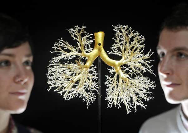
One of more than 40 curiosities on display highlights the intricacies of the human lung.
It was created by injecting resin into the organ’s air passages and dipping it into acid to corrode the spongey tissue, leaving a cast of the bronchial tree behind.
Another is a wax moulding of hands and feet taken from a patient at Edinburgh Royal Infirmary around 100 years ago, showing a congenital malformation of the nails. A cast of the internal anatomy of the human head and a set of models showing the development of a rabbit’s heart are also featured, next to a bronze anatomical figure of a flayed horse dating to 1585.
Alongside them will be a life sized hologram of the human body, believed to be the largest anatomical hologram ever made, and a colourful knitted representation of the human circulatory system.
Curator Doug Stevens, a final year fine art student at Edinburgh University, who worked with staff to create the exhibition, said: “Art and anatomy are often treated as distinct subjects, yet there is a long history of collaboration between the disciplines.
Advertisement
Hide AdAdvertisement
Hide Ad“This exhibition gives visitors the opportunity to view these rare objects and appreciate the beauty of their craftsmanship.”
He concluded: “It was a fascinating and rewarding experience exploring the university’s archives.”
• Visual Dissection – The Art of Anatomy, opens tomorrow in the university’s main library gallery and runs until March 5.