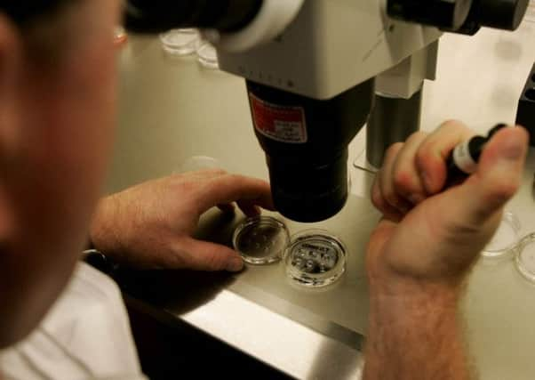
Small human ‘stomachs’ grown in lab for first time


US scientists used pluripotent stem cells, which can become any cell type in the body, to create the stomach buds, or organoids and studied infection by bugs that can cause peptic ulcers and cancer.
The tissue provides a new vehicle to fast track development of drugs as well as a model for the early stages of stomach tumours and analysing obesity-linked diabetes.
CONNECT WITH THE SCOTSMAN
Advertisement
Hide AdAdvertisement
Hide Ad• Subscribe to our daily newsletter (requires registration) and get the latest news, sport and business headlines delivered to your inbox every morning
It also is the first time researchers have produced 3D human embryonic foregut, a promising starting point for generating other early organ tissues like the lungs and pancreas.
Professor Jim Wells, of Cincinnati Children’s Hospital, said: “Until this study, no one had generated gastric cells from human pluripotent stem cells (hPSCs).
“In addition, we discovered how to promote formation of three dimensional gastric tissue with complex architecture and cellular composition.”
He added that differences between species in the embryonic development and architecture of the adult stomach make mouse models less than ideal for studying human stomach development and disease.
There are differences in the anatomy and inner workings between animals and humans, making study of disease difficult.
To create a more realistic model, Prof Wells and colleagues generated human gastric tissue using stem cells as the starting material.
The so-called gastric organoids have a complex, 3D structure and contain different cell types with functional characteristics that mirror those seen in the stomach.
Advertisement
Hide AdAdvertisement
Hide AdIn addition, the organoids are shown to faithfully recapitulate early stages of gastric disease initiated by H. pylori, demonstrating the capability of the system to act as a model to study diseases of the stomach.
Researchers can use the gastric organoids as a new way to help unlock other secrets of the stomach, such as identifying biochemical processes in the gut that allow gastric-bypass patients to become diabetes-free soon after surgery before losing significant weight.
Obesity fuelled diabetes and metabolic syndrome are an exploding public health epidemic.
Until now, a major challenge to addressing these and other medical conditions involving the stomach has been a relative lack of reliable laboratory modelling systems to accurately simulate human biology, explained Prof Wells.
The key to growing human gastric organoids was to identify the steps involved in normal stomach formation during embryonic development.
By manipulating these normal processes in a lab dish, the scientists were able to coax pluripotent stem cells toward becoming stomach.
Over the course of a month, these steps resulted in the formation of 3D human gastric organoids that were about 3mm (1/1Oth of an inch) in diameter.
The researchers whose breakthrough is reported in Nature also used this approach to identify what drives normal stomach formation in humans with the goal of understanding what goes wrong when the stomach does not form correctly.
Advertisement
Hide AdAdvertisement
Hide AdThey were surprised by how rapidly H. pylori bacteria infected stomach epithelial tissues. Within 24 hours, the bacteria had triggered biochemical changes to the organ.
The human buds faithfully mimicked the early stages of gastric disease caused by the bacteria, including the activation of a cancer gene called c-Met and the rapid spread of infection in epithelial tissues.
Another significant part of the challenge has been the relative lack of previous research literature on how the human stomach develops.
Prof Wells said they had to use a combination of published work, as well as studies from his own lab, to answer a number of basic developmental questions about its formation.
Over the course of two years, this approach of experimenting with different factors eventually resulted in the formation of 3D human gastric tissues in the petri dish.
Added Prof Wells: “This milestone would not have been possible if it had not been for previous studies from many other basic researchers on understanding embryonic organ development.”
Three-dimensional human gastric tissues made from human stem cells, described in Nature this week, provide a new system for studying human stomach development and disease.
Gastric diseases, such as ulcers and gastric cancer, affect 10 per cent of the world’s population, and their development has been associated with chronic Helicobacter pylori infection.
SEE ALSO
SCOTSMAN TABLET AND IPHONE APPS